
Gastroshiza, commonly referred to as gastroschisis, is a congenital defect of the abdominal wall that affects newborns. In this condition, the abdominal wall fails to form completely, leaving an opening through which the intestines—and sometimes other organs—protrude outside the body. Unlike omphalocele, another abdominal wall defect, in gastroshiza, the organs are not covered by a protective sac, leaving them directly exposed to amniotic fluid during pregnancy.
This exposure can cause swelling, irritation, and damage to the intestines even before birth. While the condition is alarming at first glance, medical advances in prenatal detection, neonatal care, and pediatric surgery have dramatically improved survival rates, which now exceed 90% in developed healthcare systems.
Embryology and Pathophysiology of Gastroshiza
Normal Abdominal Wall Development
During early fetal development, specifically between the 4th and 10th weeks of gestation, the embryo undergoes folding processes that shape the abdomen. The lateral body walls gradually move toward the midline and fuse to form a complete anterior abdominal wall.
At the same time, the rapidly growing intestines temporarily herniate into the umbilical cord because there isn’t enough space in the abdomen. By the 10th to 12th week, the intestines normally return to the abdominal cavity. This process requires precise coordination of tissue growth, blood supply, and mesodermal development.
Mechanism of Defect Formation
Gastroshiza arises when this closure process fails. The result is a small defect, typically on the right side of the umbilicus, through which the intestines protrude. Several theories explain this defect:
- Vascular disruption theory: Interruption of blood supply to the abdominal wall during development causes tissue necrosis and failure of closure.
- Incomplete mesodermal development: A failure in forming the mesodermal tissues that provide structural support for the abdominal wall.
- Right umbilical vein involution: Early degeneration of the right umbilical vein may weaken the adjacent tissue, increasing susceptibility to defects.
These mechanisms explain why the intestines are exposed without a protective sac, and why the defect is almost always located to the right of the belly button.
Pathophysiological Consequences of Gastroshiza
Because the intestines are directly exposed to amniotic fluid, several harmful processes occur:
- Inflammation and thickening: The fluid irritates the bowel lining, leading to edema and swelling.
- Reduced motility: Exposed intestines may have decreased movement after birth, complicating feeding.
- Increased infection risk: The exposed tissue is more prone to bacterial contamination.
- Potential malabsorption: If the bowel is damaged, nutrient absorption may be compromised.
These consequences influence the baby’s care both before and after birth.
Epidemiology and Risk Factors
Global and Regional Statistics
Gastroshiza is relatively rare, with an incidence of 2–5 per 10,000 live births globally. Interestingly, rates have been increasing over the past few decades, particularly among teenage mothers. This rise has prompted research into environmental, lifestyle, and demographic factors.
Maternal Risk Factors
Research identifies several maternal characteristics that increase risk:
- Young maternal age (<20 years): The most consistent risk factor, likely related to developmental and vascular vulnerabilities.
- Smoking, alcohol, and drug use: Substances may disrupt fetal development and blood supply.
- Poor nutrition: Deficiencies in essential vitamins and minerals can impair fetal tissue formation.
- Low BMI: Underweight mothers have a higher observed risk of delivering babies with Gastroshiza.
Although these factors increase the likelihood, many babies are born with gastroschisis in mothers without any identifiable risk factors.
Environmental and Genetic Influences
Unlike some birth defects, genetic inheritance is minimal, and the condition is considered mostly sporadic. Environmental exposures—such as certain medications, toxins, or infections—may contribute, but no definitive environmental cause has been identified.
Clinical Presentation of Gastroshiza
Prenatal Findings
Gastroshiza is often detected during routine prenatal ultrasounds, usually in the second trimester:
- Floating bowel loops: Visualized outside the fetal abdomen.
- No covering membrane: Distinguishes it from an omphalocele.
- Elevated maternal alpha-fetoprotein (AFP): Indicates leakage of fetal proteins into maternal blood.
Early detection allows healthcare providers to plan delivery in a specialized center, improving outcomes.
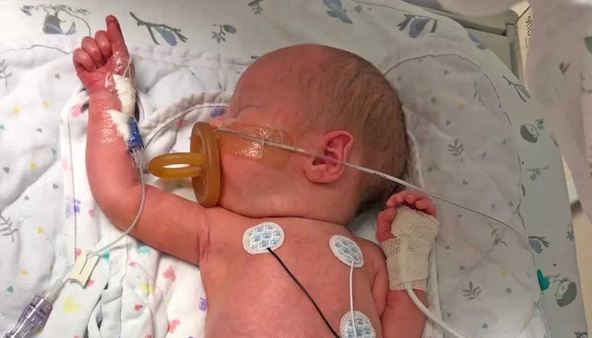
Postnatal Presentation
Immediately after birth, the condition is evident:
- Exposed intestines: Usually protruding through a small defect to the right of the umbilicus.
- Swollen, edematous bowel loops: The intestines may appear thickened or inflamed.
- Low birth weight: Common among affected newborns.
- Feeding difficulties: Often due to reduced intestinal motility and inflammation.
Prompt stabilization is crucial to prevent dehydration, infection, and heat loss.
Diagnostic Evaluation of Gastroshiza
Prenatal Diagnosis
Ultrasound is the primary tool, allowing visualization of bowel loops floating in amniotic fluid. In complex cases, fetal MRI may provide additional detail about bowel condition, associated anomalies, or abdominal cavity size.
Differential Diagnosis
Gastroshiza must be distinguished from other abdominal wall defects:
- Omphalocele: Covered sac, often associated with genetic syndromes.
- Body stalk anomaly: Severe defect involving multiple organs and connective tissue.
Accurate differentiation is critical for prognosis and surgical planning.
Postnatal Assessment of Gastroshiza
After birth, clinicians evaluate:
- Bowel viability: Color, perfusion, and integrity.
- Size of defect: Determines whether primary or staged closure is feasible.
- Associated complications: Atresia, necrosis, or perforation.
Classification and Severity Assessment
Simple Gastroshiza
- No significant intestinal complications.
- Bowel is viable and functional.
- Prognosis is excellent, and surgical recovery is generally quick.
Complex
- Includes intestinal atresia, necrosis, or perforation.
- Requires more extensive surgical intervention.
- Recovery may involve prolonged hospitalization, TPN, and close follow-up.
Severity assessment is important for prognosis and family counseling.
Prenatal Management of Gastroshiza
Monitoring During Pregnancy
- Serial ultrasounds monitor bowel dilation, edema, and growth.
- Fetal growth is tracked because intrauterine growth restriction (IUGR) may occur.
- Amniotic fluid levels are observed, as abnormal volumes may indicate bowel compromise.
Delivery Planning
- Timing: Often scheduled at 37–38 weeks to minimize complications.
- Location: Delivery in a hospital with neonatal surgery and intensive care ensures rapid intervention.
- Mode of delivery: Vaginal birth is possible in many cases; cesarean is reserved for obstetric indications rather than the condition itself.
Multidisciplinary Approach of Gastroshiza
Effective management involves coordination between:
- Obstetricians
- Neonatologists
- Pediatric surgeons
This approach ensures seamless prenatal-to-postnatal care.
Neonatal Management
Immediate Postnatal Care
Immediately after birth, babies with Gastroshiza require careful stabilization. The exposed intestines are covered with sterile, moist dressings to prevent infection and tissue damage, body temperature is maintained using incubators to avoid hypothermia, and intravenous fluids are given to prevent dehydration and maintain circulation while preparing for surgery.
Primary Closure
Primary closure is a surgical technique used when the intestines are not severely swollen or damaged. In this procedure, the exposed bowel is carefully returned to the abdominal cavity in a single operation, and the abdominal wall defect is closed, allowing faster recovery and reducing the risk of complications
Staged Closure (Silo Technique) in Gastroshiza
Staged closure, or the silo technique, is used when the intestines are too large or inflamed to fit into the abdomen immediately. A sterile silo pouch is placed over the exposed bowel, which gradually guides the intestines back into the abdominal cavity over several days. Once the bowel has returned, the defect is surgically closed.
Intensive Care Support
After surgery, babies often need specialized intensive care. Total parenteral nutrition (TPN) provides essential nutrients while the intestines recover. Ventilator support may be necessary if breathing is compromised, and antibiotics are administered to reduce the risk of infection from the exposed bowel and surgical interventions.
Complications in Gastroshiza
Short-Term Complications
- Sepsis: High risk due to exposed bowel.
- Bowel ischemia: Damage from reduced blood supply or swelling.
- Feeding intolerance: Delayed intestinal function may prolong TPN.
Long-Term Complications
- Short bowel syndrome: If part of the bowel is damaged or removed.
- Growth delays: Nutritional deficits during recovery can slow growth.
- Developmental challenges: Rare, but may require long-term monitoring.
Early intervention and meticulous care reduce these risks.
Prognosis and Survival Rates
Survival has improved dramatically:
- >90% survival in developed care systems.
- Babies with simple Gastroshiza usually recover fully and thrive.
- Prognosis depends on:
- Birth weight
- Severity of bowel damage
- Timeliness and quality of surgical care
Long-term follow-up ensures proper growth and development.
Advances in Medical and Surgical Care
Innovations in Neonatal Surgery
- Improved silo techniques reduce trauma to the intestines.
- Minimally invasive approaches decrease recovery time.
- Better materials protect exposed organs during staged repairs.
Role of NICU Improvements in Gastroshiza
- Enhanced infection control prevents sepsis.
- Precise nutrition management supports bowel healing and growth.
- Continuous monitoring allows early detection of complications.
Future Research Directions
- Identification of genetic and environmental triggers.
- Development of prenatal interventions.
- Long-term studies on quality of life and developmental outcomes.
Prevention and Public Health Perspective
While Gastroshiza cannot always be prevented, risk reduction strategies include:
- Avoiding smoking, alcohol, and harmful drugs during pregnancy.
- Maintaining adequate nutrition and taking prenatal vitamins.
- Regular prenatal checkups for early detection.
Education programs for young mothers are particularly valuable in reducing incidence.
Ethical and Psychological Considerations
A Gastroshiza diagnosis can be emotionally challenging for families.
Parental Counseling
- Families should receive clear explanations of risks, treatment options, and prognosis.
- Emotional support is essential to reduce anxiety and stress.
Decision-Making Challenges
- Severe cases may require difficult decisions about surgery and long-term care.
- Families should be guided through ethical and medical considerations.
Financial and Emotional Burden
- Prolonged hospitalization can strain families financially.
- Access to support networks and counseling can improve overall well-being.
Conclusion
Gastroshiza (gastroschisis) is a complex congenital condition with significant medical implications. Early diagnosis, prenatal monitoring, prompt neonatal care, and advanced surgical intervention allow most affected babies to survive and thrive.
While the exact cause is unclear, awareness, education, and public health initiatives can help reduce risks and improve outcomes. With modern medicine, infants with gastroschisis have a promising future, and ongoing research continues to enhance survival, growth, and quality of life.
Disclaimer:
The content of this article is intended for informational and educational purposes only and should not be considered as medical advice. Gastroschisis is a complex congenital condition, and every case may differ. Always consult a qualified healthcare professional, pediatrician, or specialist for diagnosis, treatment options, and personalized medical guidance. The author and publisher are not responsible for any decisions made based on the information provided in this article.


Leave a Reply